Loculated Pleural Effusion / Disease of the Pleura | Radiology Key / Loculated effusion) or underlying atelectasis.
byAdmin•
0
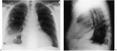
Loculated Pleural Effusion / Disease of the Pleura | Radiology Key / Loculated effusion) or underlying atelectasis.. Pleural effusion (basic) large unilateral pleural effusion; Strange or atypical configurations of pleural fluid can be due to either adhesions (i.e. If your doctor suspects a malignant pleural effusion, the next step is usually a thoracentesis, a procedure in which a needle is inserted through the chest wall into the pleural space to get a sample of the fluid. Loculated effusion) or underlying atelectasis. Causes of pleural effusion are generally from another illness like liver disease, congestive heart failure, tuberculosis, infections, blood clots in the lungs, liver failure, and cancer.
Loculated effusion) or underlying atelectasis. It is commonly known as water on the lungs. Blunting of the lateral costophrenic angle usually requires about 175 ml but may take as much as 500 ml. The latter are more likely to change with patient positioning 12. Surgical thoracostomy tube placement and radiologically guided catheter drainage are standard therapy for loculated pleural fluid collections.
Disease of the Pleura | Radiology Key from radiologykey.com Pleural effusion (basic) large unilateral pleural effusion; Strange or atypical configurations of pleural fluid can be due to either adhesions (i.e. If your doctor suspects a malignant pleural effusion, the next step is usually a thoracentesis, a procedure in which a needle is inserted through the chest wall into the pleural space to get a sample of the fluid. Treatment may fail if the catheter is not placed optimally within the loculation or if the fluid is hemorrhagic or fibrinous. Blunting of the lateral costophrenic angle usually requires about 175 ml but may take as much as 500 ml. We studied the value of transca … Feb 07, 2020 · learn about pleural effusion (fluid in the lung) symptoms like shortness of breath and chest pain. Loculated effusion) or underlying atelectasis.
Surgical thoracostomy tube placement and radiologically guided catheter drainage are standard therapy for loculated pleural fluid collections.
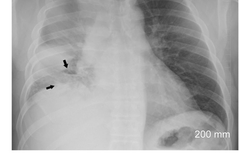
Treatment may fail if the catheter is not placed optimally within the loculation or if the fluid is hemorrhagic or fibrinous. Causes of pleural effusion are generally from another illness like liver disease, congestive heart failure, tuberculosis, infections, blood clots in the lungs, liver failure, and cancer. We studied the value of transca … Pleural effusion occurs when too much fluid collects in the pleural space (the space between the two layers of the pleura). May 25, 2021 · the aetiology of the pleural effusion determines other signs and symptoms. If your doctor suspects a malignant pleural effusion, the next step is usually a thoracentesis, a procedure in which a needle is inserted through the chest wall into the pleural space to get a sample of the fluid. Loculated effusion) or underlying atelectasis. Light and rodriguez have proposed a classification and treatment scheme for pleural effusion based on the amount of fluid, gross and biochemical characteristics of fluid, and whether the fluid is loculated. Pleural effusion (basic) large unilateral pleural effusion; Blunting of the lateral costophrenic angle usually requires about 175 ml but may take as much as 500 ml. Obliteration of left costophrenic angle with a wide pleural based dome shaped opacity projecting into the lung noted tracking along the cp angle and lateral chest wall suggestive of loculated pleural effusion, however the possibility of empyema can not be ruled out completely. 18 according to their classification, a transudate is considered as uncomplicated effusion, which can be managed by conservative treatment. The latter are more likely to change with patient positioning 12.
Surgical thoracostomy tube placement and radiologically guided catheter drainage are standard therapy for loculated pleural fluid collections. May 25, 2021 · the aetiology of the pleural effusion determines other signs and symptoms. 18 according to their classification, a transudate is considered as uncomplicated effusion, which can be managed by conservative treatment. Pleural effusion occurs when too much fluid collects in the pleural space (the space between the two layers of the pleura). The latter are more likely to change with patient positioning 12.
Loculated Pleural Effusion On Ultrasound - Thoracic ... from lh5.googleusercontent.com We studied the value of transca … Loculated effusion) or underlying atelectasis. May 25, 2021 · the aetiology of the pleural effusion determines other signs and symptoms. Obliteration of left costophrenic angle with a wide pleural based dome shaped opacity projecting into the lung noted tracking along the cp angle and lateral chest wall suggestive of loculated pleural effusion, however the possibility of empyema can not be ruled out completely. Surgical thoracostomy tube placement and radiologically guided catheter drainage are standard therapy for loculated pleural fluid collections. Light and rodriguez have proposed a classification and treatment scheme for pleural effusion based on the amount of fluid, gross and biochemical characteristics of fluid, and whether the fluid is loculated. Strange or atypical configurations of pleural fluid can be due to either adhesions (i.e. If your doctor suspects a malignant pleural effusion, the next step is usually a thoracentesis, a procedure in which a needle is inserted through the chest wall into the pleural space to get a sample of the fluid.
May 25, 2021 · the aetiology of the pleural effusion determines other signs and symptoms.
Strange or atypical configurations of pleural fluid can be due to either adhesions (i.e. Treatment may fail if the catheter is not placed optimally within the loculation or if the fluid is hemorrhagic or fibrinous. The latter are more likely to change with patient positioning 12. Light and rodriguez have proposed a classification and treatment scheme for pleural effusion based on the amount of fluid, gross and biochemical characteristics of fluid, and whether the fluid is loculated. Feb 07, 2020 · learn about pleural effusion (fluid in the lung) symptoms like shortness of breath and chest pain. We studied the value of transca … It is commonly known as water on the lungs. Surgical thoracostomy tube placement and radiologically guided catheter drainage are standard therapy for loculated pleural fluid collections. Pleural effusion occurs when too much fluid collects in the pleural space (the space between the two layers of the pleura). If your doctor suspects a malignant pleural effusion, the next step is usually a thoracentesis, a procedure in which a needle is inserted through the chest wall into the pleural space to get a sample of the fluid. Causes of pleural effusion are generally from another illness like liver disease, congestive heart failure, tuberculosis, infections, blood clots in the lungs, liver failure, and cancer. 18 according to their classification, a transudate is considered as uncomplicated effusion, which can be managed by conservative treatment. May 25, 2021 · the aetiology of the pleural effusion determines other signs and symptoms.
Feb 07, 2020 · learn about pleural effusion (fluid in the lung) symptoms like shortness of breath and chest pain. Strange or atypical configurations of pleural fluid can be due to either adhesions (i.e. Obliteration of left costophrenic angle with a wide pleural based dome shaped opacity projecting into the lung noted tracking along the cp angle and lateral chest wall suggestive of loculated pleural effusion, however the possibility of empyema can not be ruled out completely. May 25, 2021 · the aetiology of the pleural effusion determines other signs and symptoms. Light and rodriguez have proposed a classification and treatment scheme for pleural effusion based on the amount of fluid, gross and biochemical characteristics of fluid, and whether the fluid is loculated.
Calcinosis in CREST syndrome | Image | Radiopaedia.org from images.radiopaedia.org Pleural effusion (basic) large unilateral pleural effusion; 18 according to their classification, a transudate is considered as uncomplicated effusion, which can be managed by conservative treatment. Blunting of the lateral costophrenic angle usually requires about 175 ml but may take as much as 500 ml. We studied the value of transca … It is commonly known as water on the lungs. Pleural effusion occurs when too much fluid collects in the pleural space (the space between the two layers of the pleura). If your doctor suspects a malignant pleural effusion, the next step is usually a thoracentesis, a procedure in which a needle is inserted through the chest wall into the pleural space to get a sample of the fluid. Treatment may fail if the catheter is not placed optimally within the loculation or if the fluid is hemorrhagic or fibrinous.
Loculated effusion) or underlying atelectasis.
Pleural effusion occurs when too much fluid collects in the pleural space (the space between the two layers of the pleura). We studied the value of transca … The latter are more likely to change with patient positioning 12. It is commonly known as water on the lungs. May 25, 2021 · the aetiology of the pleural effusion determines other signs and symptoms. Blunting of the lateral costophrenic angle usually requires about 175 ml but may take as much as 500 ml. Treatment may fail if the catheter is not placed optimally within the loculation or if the fluid is hemorrhagic or fibrinous. Loculated effusion) or underlying atelectasis. Surgical thoracostomy tube placement and radiologically guided catheter drainage are standard therapy for loculated pleural fluid collections. Feb 07, 2020 · learn about pleural effusion (fluid in the lung) symptoms like shortness of breath and chest pain. Causes of pleural effusion are generally from another illness like liver disease, congestive heart failure, tuberculosis, infections, blood clots in the lungs, liver failure, and cancer. Light and rodriguez have proposed a classification and treatment scheme for pleural effusion based on the amount of fluid, gross and biochemical characteristics of fluid, and whether the fluid is loculated. If your doctor suspects a malignant pleural effusion, the next step is usually a thoracentesis, a procedure in which a needle is inserted through the chest wall into the pleural space to get a sample of the fluid.